
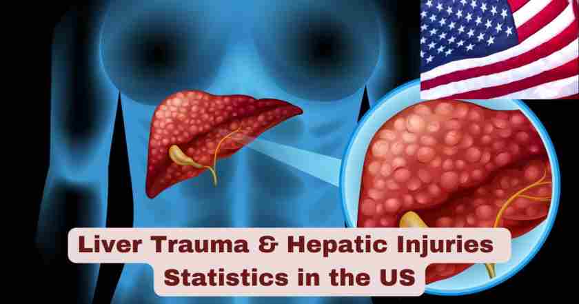
Liver Trauma and Hepatic Injuries in the US 2025
Liver trauma represents one of the most critical and life-threatening conditions encountered in emergency medicine across the United States. As the largest solid organ in the abdomen, the liver’s relatively fixed anatomical position makes it particularly vulnerable to both blunt and penetrating injuries. When traumatic forces impact the abdomen, the liver frequently bears the brunt of the damage, leading to a spectrum of injuries ranging from minor capsular tears to devastating parenchymal disruptions with massive hemorrhage. The management of hepatic injuries has evolved dramatically over the past several decades, with modern trauma centers now employing sophisticated diagnostic tools and treatment protocols that have significantly improved survival rates for patients experiencing these potentially fatal injuries.
The clinical significance of liver trauma extends far beyond individual patient outcomes, representing a substantial burden on the American healthcare system. These injuries predominantly affect young adults during their most productive years, resulting in significant economic costs related to emergency care, surgical interventions, intensive care unit stays, and long-term rehabilitation. Understanding the epidemiology, patterns, and outcomes of hepatic trauma in the US 2025 is essential for healthcare providers, policymakers, and trauma system administrators working to optimize care delivery and resource allocation. Recent advances in non-operative management strategies, interventional radiology techniques, and damage control surgery have transformed how clinicians approach these complex injuries, though mortality rates remain concerning for severe grades of liver trauma.
Interesting Facts and Latest Statistics on Liver Trauma in the US 2025
| Key Fact Category | Statistical Data | Clinical Significance |
|---|---|---|
| Incidence Among Abdominal Injuries | 22% of all abdominal trauma cases involve liver injuries | Liver is second most commonly injured abdominal organ after spleen |
| Minor Injury Prevalence | 80-90% of hepatic trauma cases are Grade I or II injuries | Most liver injuries are relatively minor and manageable non-operatively |
| Non-Operative Management Success | Over 80% of liver injuries treated without surgery | Shift toward conservative management has improved outcomes |
| Overall Mortality Rate | 8% average mortality (range 4.1-11.7%) | Significant death risk despite advances in trauma care |
| Grade Distribution in Recent Data | 60% Grade I/II, 21% Grade III, 14% Grade IV, 5% Grade V, 0.2% Grade VI | Higher grades correlate with exponentially increased mortality |
| Penetrating vs Blunt Mechanism | 24% penetrating injuries, 74% blunt trauma cases | Blunt mechanisms dominate in civilian trauma settings |
| High-Grade Injury Complications | 15-20% complication rate for high-grade injuries | Serious injuries carry significant morbidity beyond mortality |
| Primary Cause of Death | Liver injury is leading cause of death in severe abdominal trauma | Hemorrhage from hepatic vessels drives fatal outcomes |
| Hemodynamic Instability Factor | Treatment decisions rely on stability rather than injury grade alone | Patient physiology guides management more than anatomy |
| Embolization Mortality Rate | 10% mortality rate for patients requiring arterial embolization | Interventional procedures offer life-saving option for bleeding |
Data Sources: National Trauma Data Bank (NTDB) – American College of Surgeons, NCBI StatPearls Liver Trauma Reference, American Association for the Surgery of Trauma (AAST) Organ Injury Scale studies, Trauma Quality Programs Participant Use File, Contemporary epidemiologic analysis of ACS TQIP database (2025), Radiopaedia clinical references.
The data presented reveals several critical insights about the current state of hepatic trauma management in the US. The predominance of minor liver injuries (80-90% being Grade I or II) demonstrates that while hepatic trauma is relatively common in trauma patients, most cases involve limited tissue damage that can be successfully managed through observation and supportive care. This finding has significant implications for trauma system resource allocation, as it suggests that the majority of liver trauma patients can be cared for effectively without immediate surgical intervention. The 22% incidence rate among all abdominal injuries underscores the liver’s vulnerability during traumatic events, reinforcing the need for rapid diagnostic imaging and experienced trauma team evaluation for any patient with suspected abdominal trauma.
The overall mortality rate of 8% associated with liver injuries represents a sobering reality despite decades of advances in trauma care protocols. This mortality figure, with its range from 4.1% to 11.7% across different trauma centers and patient populations, reflects the inherent danger of hepatic vascular injuries and the challenges of managing severe hemorrhage in critically injured patients. The dramatic shift toward non-operative management (NOM) succeeding in over 80% of cases represents one of the most significant paradigm changes in trauma surgery over the past generation. This conservative approach, guided by hemodynamic stability and serial clinical assessments rather than injury grade alone, has reduced the need for emergency laparotomy and its associated complications. The distribution data showing 60% of injuries classified as Grade I/II but only 0.2% as Grade VI illustrates the severity spectrum, with the rarest injuries being virtually uniformly fatal. These statistics collectively paint a picture of liver trauma in 2025 as a condition where most patients survive with appropriate care, but severe injuries continue to challenge even the most advanced trauma systems in the United States.
Liver Trauma Incidence and Epidemiology in the US 2025
| Epidemiological Factor | Statistical Finding | Population Impact |
|---|---|---|
| Total Cases Analyzed (Recent Multi-Year Data) | 96,652 patients with liver trauma identified in national database | Large-scale data provides robust epidemiological insights |
| Patients Included After Criteria | 60,199 patients met inclusion criteria for detailed analysis | Represents comprehensive contemporary US experience |
| Penetrating Mechanism | 24% of all cases resulted from penetrating trauma | Gunshot wounds and stabbings comprise minority |
| Blunt Mechanism | 74% of all cases resulted from blunt force trauma | Motor vehicle crashes and falls dominate causation |
| Age Group Most Affected | Young adults aged 18-45 years represent majority of cases | Trauma strikes during peak productive life years |
| Gender Distribution | Male patients comprise approximately 70% of liver trauma cases | Males experience disproportionate trauma burden |
| Urban vs Rural Patterns | Higher penetrating trauma rates in urban trauma centers | Geographic location influences injury mechanism patterns |
| Seasonal Variation | Summer months show 15-20% increase in trauma admissions | Warmer weather correlates with higher injury rates |
| Associated Injuries | 65-75% of liver trauma patients have concurrent injuries | Isolated hepatic trauma is relatively uncommon |
| Interfacility Transfers | Significant proportion require transfer to higher-level centers | Complex cases need specialized trauma center resources |
Data Sources: Contemporary epidemiologic overview from American College of Surgeons Trauma Quality Improvement Program (TQIP) database analysis (2025), National Trauma Data Bank annual reports, Coalition for National Trauma Research statistics.
The epidemiological landscape of liver trauma in the United States in 2025 reveals important patterns that inform trauma system planning and resource allocation decisions. The substantial dataset of 96,652 patients identified with hepatic injuries across participating trauma centers demonstrates the scope of this clinical challenge facing American healthcare. With 60,199 patients meeting rigorous inclusion criteria for detailed analysis, researchers have unprecedented insight into the contemporary patterns of liver injury across diverse geographic regions and trauma center levels. The 74% blunt trauma versus 24% penetrating trauma distribution reflects the dominance of motor vehicle collisions, motorcycle crashes, falls from height, and assault-related mechanisms in civilian trauma populations. This ratio varies significantly between urban trauma centers serving areas with higher violent crime rates and rural facilities where agricultural and recreational accidents predominate.
The demographic patterns revealed in these statistics highlight that liver trauma disproportionately affects young male adults during their most economically productive years. The concentration of cases among individuals aged 18-45 years and the approximate 70% male predominance mirrors broader trauma epidemiology patterns. This demographic reality translates into enormous societal costs beyond immediate medical expenses, including lost productivity, disability, and family impact. The finding that 65-75% of liver trauma patients present with associated injuries emphasizes the complex, multi-system nature of serious traumatic events. Patients rarely sustain isolated hepatic trauma; rather, liver injuries typically occur within the context of polytrauma involving the head, chest, extremities, or other abdominal organs. This constellation of injuries complicates clinical decision-making and necessitates coordinated multidisciplinary care approaches. The seasonal variation showing 15-20% increases during summer months reflects patterns of increased outdoor activity, travel, and recreational pursuits during warmer weather. Understanding these epidemiological patterns of liver trauma in the US in 2025 enables trauma systems to anticipate resource needs and optimize staffing patterns to meet predictable fluctuations in patient volume.
Liver Injury Grading and Severity Classification in the US 2025
| AAST Injury Grade | Percentage Distribution | Anatomical Description | Mortality Risk |
|---|---|---|---|
| Grade I | 30-35% of all cases | Subcapsular hematoma <10% surface area, capsular tear <1cm depth | <3% mortality |
| Grade II | 25-30% of all cases | Subcapsular hematoma 10-50% surface area, laceration 1-3cm depth | 3-5% mortality |
| Grade III | 21% of all cases | Subcapsular hematoma >50% surface area, laceration >3cm depth | 10-15% mortality |
| Grade IV | 14% of all cases | Parenchymal disruption 25-75% of hepatic lobe, active bleeding | 25-40% mortality |
| Grade V | 5% of all cases | Parenchymal disruption >75% of lobe, juxtahepatic venous injury | 50-70% mortality |
| Grade VI | 0.2% of all cases | Hepatic avulsion, complete vascular devascularization | >80% mortality (often fatal) |
| Grades I-II Combined | 60% of total liver injuries | Minor hepatic trauma amenable to non-operative management | Excellent prognosis overall |
| Grades IV-V Combined | 19% of total cases | Major hepatic trauma requiring aggressive intervention | High mortality despite treatment |
| Vascular Injury Component | Incorporated in 2018 AAST-OIS revision | Active contrast extravasation, pseudoaneurysm, AV fistula | Significantly increases complication risk |
| Biliary Injury Component | Present in 5-10% of cases | Bile duct disruption, biloma formation | Requires specialized intervention |
Data Sources: American Association for the Surgery of Trauma (AAST) Organ Injury Scale 2018 revision, Contemporary TQIP database analysis (2025), NCBI StatPearls clinical references, World Society of Emergency Surgery guidelines.
The American Association for the Surgery of Trauma Organ Injury Scale (AAST-OIS) provides the standardized framework for classifying hepatic trauma severity across trauma centers nationwide. This grading system, most recently revised in 2018, incorporates both anatomical injury extent and vascular injury patterns identified on computed tomography imaging. The data showing that 60% of all liver injuries fall into the Grade I or II categories provides reassurance that most patients with hepatic trauma have relatively minor injuries that can be successfully managed through non-operative approaches. These lower-grade injuries typically involve limited capsular tears or small subcapsular hematomas that pose minimal risk of ongoing hemorrhage or hemodynamic compromise. The associated mortality rates under 5% for these minor injuries reflect the generally favorable prognosis when patients remain hemodynamically stable and can be monitored closely in an appropriate care setting.
The statistical distribution across injury grades reveals a clear inverse relationship between injury severity and frequency. While Grade I and II injuries comprise 60% of cases, the most severe Grade VI injuries represent only 0.2% of the total. However, this rarity should not minimize their clinical significance, as Grade VI hepatic injuries involving complete hepatic avulsion or juxtahepatic venous disruption carry mortality rates exceeding 80% and often result in death before patients reach definitive care. The Grade III injuries (21% of cases) represent a transitional category where clinical decision-making becomes more nuanced. These moderate injuries involving lacerations exceeding 3cm depth or large subcapsular hematomas require careful assessment and typically necessitate admission to intensive care settings with serial hemoglobin monitoring and repeat imaging. The combined 19% of patients presenting with Grade IV or V injuries face substantially elevated mortality risk in the 25-70% range, reflecting the massive parenchymal disruption and major vascular injuries that characterize these severe trauma patterns. The incorporation of vascular injury findings in the 2018 AAST-OIS revision acknowledges that active contrast extravasation, pseudoaneurysm formation, and arteriovenous fistulas significantly impact clinical outcomes regardless of parenchymal injury extent. Understanding these liver injury grades and their associated outcomes in the US in 2025 enables trauma surgeons to communicate prognosis, guide treatment decisions, and establish appropriate monitoring protocols for diverse patient presentations.
Liver Trauma Mortality and Survival Outcomes in the US 2025
| Outcome Metric | Statistical Data | Clinical Context |
|---|---|---|
| Overall Mortality Rate | 8% (range 4.1-11.7% across studies) | Significant variation based on injury severity and center experience |
| Grade I Mortality | <3% death rate | Excellent survival for minor injuries |
| Grade II Mortality | 3-5% death rate | Low mortality with appropriate monitoring |
| Grade III Mortality | 10-15% death rate | Moderate risk requiring ICU-level care |
| Grade IV Mortality | 25-40% death rate | High mortality despite aggressive intervention |
| Grade V Mortality | 50-70% death rate | Majority of patients with severe injuries die |
| Grade VI Mortality | >80% death rate | Often fatal before reaching hospital |
| Non-Operative Management Survival | >92% survival rate for NOM patients | Conservative approach succeeds in hemodynamically stable patients |
| Surgical Intervention Mortality | 15-25% mortality for operative cases | Surgery reserved for unstable or deteriorating patients |
| Arterial Embolization Mortality | 10% death rate for embolized patients | Interventional radiology offers middle-ground option |
Data Sources: National Trauma Data Bank outcomes data, StatPearls Liver Trauma mortality statistics, Hepatic trauma interventions studies (PMC), Radiopaedia clinical outcome references, TQIP database survival analysis.
The mortality statistics for liver trauma in the United States demonstrate the critical importance of injury severity grading in predicting patient outcomes. The overall mortality rate of 8% represents an aggregate figure encompassing the entire spectrum from minor subcapsular hematomas to devastating hepatic avulsions, but this single number obscures the dramatic variation in death risk across injury grades. The range of 4.1-11.7% across different studies and trauma centers reflects institutional differences in patient populations, transfer patterns, resource availability, and clinical expertise. Level I trauma centers serving as regional referral hubs typically receive a higher proportion of severe injuries transferred from outlying facilities, which can elevate their mortality statistics despite providing excellent care. Conversely, community trauma centers managing predominantly lower-grade injuries may report mortality rates at the lower end of the spectrum.
The progressive escalation in mortality risk from under 3% for Grade I injuries to over 80% for Grade VI injuries illustrates the exponential increase in death risk as injury severity advances through the classification scale. This pattern underscores why the AAST grading system serves not merely as an anatomical classification but as a powerful prognostic tool. The 92% survival rate achieved with non-operative management represents one of the great success stories in modern trauma care. Over the past three decades, the trauma surgery community has progressively expanded indications for conservative management, demonstrating that hemodynamically stable patients with even relatively significant hepatic lacerations can often be safely managed through observation, serial hemoglobin monitoring, and repeat imaging rather than immediate surgical exploration. This paradigm shift has reduced the complications associated with laparotomy, including surgical site infections, abdominal compartment syndrome, and the physiological stress of major surgery in already compromised patients.
The 15-25% mortality rate for patients requiring surgical intervention reflects the fact that operative management is now largely reserved for those who demonstrate hemodynamic instability or ongoing hemorrhage that cannot be controlled through other means. These patients, by definition, represent a higher-risk cohort with more severe injuries and greater physiological derangement. The 10% mortality rate for patients undergoing arterial embolization positions this interventional radiology technique as an important middle option between observation and surgery. When imaging identifies active contrast extravasation indicating ongoing bleeding, angiography with selective embolization of bleeding hepatic arterial branches can achieve hemostasis without the morbidity of laparotomy. Understanding these mortality and survival outcomes for hepatic trauma in 2025 enables trauma teams to counsel families realistically about prognosis while recognizing that modern trauma care has significantly improved survival rates compared to historical outcomes, particularly for patients who reach definitive care with survivable injuries and adequate physiological reserve.
Treatment Approaches and Management Strategies for Liver Trauma in the US 2025
| Management Strategy | Utilization Rate | Indications and Applications | Success Rate |
|---|---|---|---|
| Non-Operative Management (NOM) | >80% of all liver injuries | Hemodynamically stable patients, Grades I-IV, no peritonitis | >92% success rate |
| Serial Clinical Assessment | Standard protocol in 100% of NOM cases | Hourly vital signs, serial abdominal exams, hemoglobin monitoring | Critical for early detection of failure |
| Repeat CT Imaging | Performed in 40-50% of NOM patients | Scheduled follow-up or clinical deterioration | Identifies delayed bleeding or complications |
| Angiography with Embolization | 8-12% of liver trauma cases | Active contrast extravasation on CT, Grade IV-V injuries | 85-90% hemostasis achieved |
| Damage Control Surgery | 10-15% of severe cases | Hemodynamic instability, hemorrhagic shock, coagulopathy | Life-saving for unstable patients |
| Definitive Surgical Repair | 5-8% of total cases | Failed NOM, ongoing bleeding despite embolization | Reserved for refractory hemorrhage |
| Hepatic Resection | <2% of cases | Completely devascularized tissue, uncontrollable bleeding | High-risk procedure of last resort |
| Perihepatic Packing | Used in 60-70% of damage control cases | Temporary hemorrhage control, allows resuscitation | Requires staged reoperation |
| Blood Product Transfusion | Required in 30-40% of admissions | Hemoglobin <7-8 g/dL, ongoing hemorrhage, shock | Massive transfusion protocol for severe cases |
| ICU Admission | 70-80% of Grade III-V injuries | Hemodynamic monitoring, serial labs, intervention capability | Standard of care for moderate-severe injuries |
Data Sources: World Society of Emergency Surgery (WSES) 2020 liver trauma guidelines, Hepatic trauma interventions PMC studies, Contemporary management analysis from TQIP database, MSD Manual hepatic injury treatment protocols, NCBI clinical management reviews.
The contemporary management of liver trauma in the United States in 2025 reflects a sophisticated, algorithm-driven approach that prioritizes patient physiology over anatomical injury grade in making treatment decisions. The dominant paradigm of non-operative management (NOM) succeeding in over 80% of cases represents a fundamental transformation from the surgical approach that prevailed through the 1980s, when most significant hepatic injuries prompted exploratory laparotomy. Modern trauma protocols recognize that the liver possesses remarkable intrinsic hemostatic capabilities, with minor bleeding often stopping spontaneously as thrombosis occurs within injured vessels and parenchyma. The greater than 92% success rate with NOM validates this conservative approach, demonstrating that carefully selected patients can be safely managed through observation alone. The key criterion for NOM candidacy is hemodynamic stability, defined as maintaining adequate blood pressure and perfusion without requiring massive ongoing transfusion support.
The protocol of serial clinical assessment employed in all NOM cases provides the safety net that enables conservative management. Patients undergo hourly vital sign monitoring, frequent abdominal examinations by experienced clinicians, and serial hemoglobin measurements to detect occult ongoing bleeding. Any sign of clinical deterioration, including increasing abdominal distension, peritoneal signs, persistent tachycardia, dropping hemoglobin despite transfusion, or hemodynamic instability, prompts immediate reassessment and potential escalation to interventional or surgical management. Repeat CT imaging performed in 40-50% of NOM patients serves to identify delayed complications such as pseudoaneurysm formation, expanding hematomas, bile leaks, or the development of intraperitoneal fluid collections that might indicate ongoing hemorrhage or bile peritonitis.
Angiography with selective arterial embolization has emerged as a critical intermediate intervention, utilized in 8-12% of liver trauma cases. When initial or follow-up CT imaging demonstrates active contrast extravasation, indicating ongoing arterial bleeding, patients can be taken to the interventional radiology suite for diagnostic angiography. Once bleeding vessels are identified, they can be selectively embolized using coils, gelfoam, or other occlusive agents, achieving hemostasis in 85-90% of cases without the need for laparotomy. This technique is particularly valuable for Grade IV and V injuries where the risk of NOM failure is higher but patients remain stable enough to tolerate the time required for angiographic intervention.
For the 10-15% of severe cases presenting with hemorrhagic shock, profound coagulopathy, or hemodynamic instability despite aggressive resuscitation, damage control surgery represents the life-saving intervention. This approach prioritizes rapid hemorrhage control through techniques such as perihepatic packing (used in 60-70% of damage control cases), where surgical packs are placed around the liver to achieve tamponade of bleeding surfaces. Rather than attempting definitive repair during the initial operation when the patient is physiologically depleted, surgeons focus on achieving temporary hemostasis, closing the abdomen (sometimes leaving it open if abdominal compartment syndrome is a concern), and transferring the patient to intensive care for aggressive resuscitation aimed at correcting hypothermia, acidosis, and coagulopathy. These patients return to the operating room 24-48 hours later for pack removal and reassessment once physiological parameters have improved. Definitive surgical repair including techniques like hepatorrhaphy, selective vascular ligation, or rarely hepatic resection (performed in under 2% of cases) is reserved for patients who fail NOM or have injuries that cannot be controlled through less invasive means. Understanding these diverse treatment approaches for liver trauma in 2025 enables trauma teams to match interventions appropriately to individual patient presentations, maximizing survival while minimizing unnecessary surgical morbidity.
Complications and Morbidity Associated with Liver Trauma in the US 2025
| Complication Type | Incidence Rate | Clinical Presentation | Management Approach |
|---|---|---|---|
| Overall Complication Rate | <7% for all grades | Varies widely based on injury severity | Higher rates with operative management |
| High-Grade Injury Complications | 15-20% for Grade IV-V | Multiple organ involvement, complex recovery | Requires prolonged hospitalization |
| Biliary Complications | 5-10% of cases | Bile leak, biloma formation, biliary peritonitis | Percutaneous drainage, ERCP with stenting |
| Hepatic Abscess | 2-3% incidence | Fever, leukocytosis, imaging findings | Percutaneous drainage, antibiotics |
| Hemobilia | 1-2% of cases | Gastrointestinal bleeding, melena, anemia | Angiographic embolization |
| Hepatic Necrosis | <5% after embolization | Post-embolization syndrome, fever, elevated LFTs | Usually self-limited, supportive care |
| Abdominal Compartment Syndrome | 3-5% of operative cases | Elevated intra-abdominal pressure, organ dysfunction | Decompressive laparotomy |
| Coagulopathy | 25-30% of severe trauma | Trauma-induced coagulopathy, massive transfusion | Aggressive blood product replacement |
| Sepsis and Infection | 8-10% of high-grade injuries | Systemic inflammatory response, multiorgan dysfunction | Broad-spectrum antibiotics, source control |
| Non-Operative Management Failure | 8% failure rate | Hemodynamic deterioration, increasing transfusion needs | Conversion to angiography or surgery |
Data Sources: MSD Manual Professional Edition hepatic injury complications, NCBI hepatic trauma complication studies, Liver trauma WSES guidelines, NTDB complication reporting trends analysis, Interventional radiology outcomes data.
The complication profile for liver trauma represents a significant source of morbidity beyond initial mortality, particularly for patients with high-grade injuries requiring aggressive intervention. The overall complication rate of less than 7% across all injury grades reflects the generally favorable outcomes achieved with modern trauma care, but this aggregate figure obscures the substantially elevated risk faced by patients with severe injuries. The dramatic increase to 15-20% complication rates for Grade IV and V injuries underscores the complex clinical course these patients often experience, with complications extending hospital stays, requiring additional interventions, and potentially contributing to long-term disability or delayed mortality.
Biliary complications, occurring in 5-10% of liver trauma cases, represent one of the most clinically significant post-injury problems. When hepatic lacerations involve the biliary tree, bile can leak into the peritoneal cavity causing chemical peritonitis, or accumulate in collections called bilomas that may become secondarily infected. These complications typically manifest several days after injury, presenting with persistent abdominal pain, fever, leukocytosis, and elevated liver enzymes. Management usually involves percutaneous drainage of bilomas under imaging guidance, with some cases requiring endoscopic retrograde cholangiopancreatography (ERCP) to identify bile duct injuries and place stents to facilitate healing. Hepatic abscess formation occurs in 2-3% of cases, typically developing as a consequence of devitalized hepatic tissue becoming infected or as a secondary infection of hematomas or bilomas. These require percutaneous drainage and prolonged antibiotic therapy, adding weeks to hospitalization and recovery time.
Hemobilia, the uncommon but characteristic complication occurring in 1-2% of liver trauma patients, results from abnormal communication between hepatic arterial vessels and the biliary tree. Patients present with the classic triad of gastrointestinal bleeding, right upper quadrant pain, and jaundice, though all three features are not always present. This complication typically develops days to weeks after injury and requires angiographic diagnosis and selective arterial embolization for treatment. Hepatic necrosis following arterial embolization, occurring in under 5% of cases, reflects the tradeoff inherent in sacrificing hepatic arterial blood flow to achieve hemostasis. Fortunately, the liver’s dual blood supply from both hepatic artery and portal vein usually provides sufficient perfusion to prevent extensive tissue death, and most post-embolization necrosis remains limited and clinically manageable with supportive care.
The 25-30% incidence of coagulopathy among patients with severe hepatic trauma reflects the phenomenon of trauma-induced coagulopathy, where the combination of tissue injury, hemorrhage, hypothermia, acidosis, and dilutional effects of massive fluid resuscitation creates a vicious cycle of impaired hemostasis. This complication necessitates aggressive blood product replacement following massive transfusion protocols that provide red blood cells, plasma, and platelets in balanced ratios to restore coagulation function. Abdominal compartment syndrome, developing in 3-5% of operative cases, occurs when increased intra-abdominal pressure from hemorrhage, tissue edema, and fluid accumulation compromises perfusion of abdominal organs and impairs ventilation. This life-threatening complication requires emergency decompressive laparotomy to relieve pressure and restore organ perfusion.
The 8% failure rate of non-operative management represents patients who initially appeared stable enough for conservative treatment but subsequently deteriorated, requiring escalation to angiographic embolization or surgical intervention. Risk factors for NOM failure include higher injury grades, presence of active contrast extravasation on initial CT, large hemoperitoneum, and need for transfusion of multiple blood units. Understanding the complication spectrum for liver trauma in the US in 2025 enables clinicians to anticipate problems, implement surveillance protocols to detect complications early, and intervene promptly when issues arise, ultimately improving long-term outcomes beyond simply achieving initial survival.
Hospital Resource Utilization and Economic Impact of Liver Trauma in the US 2025
| Resource Metric | Statistical Data | Healthcare System Impact |
|---|---|---|
| Average Hospital Length of Stay | 8-12 days for moderate-severe injuries | Substantial bed utilization in trauma centers |
| ICU Length of Stay | 3-7 days for Grade III-V injuries | Intensive resource consumption and staffing |
| Average Hospital Charges | $50,000-$150,000 per admission | Wide variation based on injury severity and complications |
| Operative Cases Cost | 2-3 times higher than NOM cases | Surgical intervention substantially increases expenses |
| Blood Product Costs | $5,000-$25,000 for severe cases | Massive transfusion protocols extremely costly |
| ICU Daily Costs | $4,000-$8,000 per day | Critical care represents major expense component |
| Readmission Rate | 8-12% within 30 days | Complications drive unplanned readmissions |
| Disability and Lost Productivity | Estimated $100,000+ per severe case | Societal costs exceed direct medical expenses |
| Trauma Center Preparation Costs | Ongoing operational expenses | 24/7 team availability requires substantial infrastructure |
| Long-Term Follow-Up Care | 15-20% require extended outpatient management | Imaging surveillance, specialty consultations |
Data Sources: CDC Economic Cost of Injury 2019 report, National Trauma Data Bank hospital utilization data, Coalition for National Trauma Research economic impact studies, Healthcare cost analysis for trauma admissions.
The economic burden of liver trauma on the United States healthcare system extends far beyond direct medical costs to encompass broader societal impacts including lost productivity, disability, rehabilitation expenses, and long-term care needs. The average hospital length of stay of 8-12 days for patients with moderate to severe hepatic injuries represents substantial bed utilization in trauma centers that must maintain capacity to receive critically injured patients at all hours. When multiplied across the tens of thousands of liver trauma admissions occurring annually nationwide, this translates into hundreds of thousands of hospital-days devoted to caring for these patients. The ICU length of stay averaging 3-7 days for Grade III-V injuries reflects the resource-intensive nature of caring for severely injured trauma patients who require continuous hemodynamic monitoring, frequent laboratory studies, potential interventions, and high nurse-to-patient ratios characteristic of intensive care settings.
The wide range of average hospital charges from $50,000 to $150,000 per admission reflects the tremendous variability in injury severity, treatment approaches, complication rates, and hospital cost structures across different facilities and geographic regions. Patients with minor Grade I or II injuries who remain stable throughout a brief observation period may incur charges at the lower end of this spectrum, while those with Grade IV or V injuries requiring damage control surgery, massive transfusion, prolonged ICU stays, and management of multiple complications can accumulate charges well into the hundreds of thousands of dollars. The finding that operative cases cost 2-3 times more than non-operative management cases provides compelling economic justification for the contemporary preference toward conservative management when clinically appropriate. Beyond direct cost savings, NOM avoids the morbidity associated with surgical complications, further reducing overall healthcare expenditures.
Blood product costs, ranging from $5,000 to $25,000 for patients requiring massive transfusion, represent another substantial expense component. The most severely injured patients may receive dozens of units of packed red blood cells, fresh frozen plasma, platelets, and cryoprecipitate during resuscitation, with each unit carrying both acquisition costs and the extensive infrastructure required for blood banking, typing, crossmatching, and safe administration. The ICU daily costs of $4,000-$8,000 multiply rapidly over the typical 3-7 day critical care stay, contributing significantly to total hospitalization expenses. The 8-12% readmission rate within 30 days adds additional costs as patients return with complications such as bilomas, abscesses, or delayed hemorrhage requiring intervention. Perhaps most significantly from a societal perspective, the estimated $100,000+ per severe case in disability and lost productivity costs reflects that liver trauma predominantly affects young adults during peak earning years. When individuals suffer permanent disability or prolonged recovery periods preventing return to work, the economic impact extends far beyond medical bills to affect families, employers, and society broadly. Understanding these economic dimensions of liver trauma in the US in 2025 provides essential context for healthcare policy decisions, trauma system funding priorities, and injury prevention initiatives aimed at reducing the incidence of these costly injuries.
Diagnostic Imaging and Technology in Liver Trauma Management in the US 2025
| Imaging Modality | Utilization Rate | Diagnostic Capabilities | Clinical Applications |
|---|---|---|---|
| Computed Tomography (CT) | >95% of hemodynamically stable patients | Gold standard for injury grading, active bleeding detection | Primary diagnostic tool in trauma evaluation |
| Focused Assessment with Sonography for Trauma (FAST) | 80-90% of initial trauma evaluations | Rapid detection of free fluid (blood) in abdomen | Bedside screening for hemodynamically unstable patients |
| Contrast-Enhanced CT | Standard protocol in 100% of suspected liver injuries | Identifies active extravasation, vascular injuries, organ lacerations | Enables accurate AAST grade classification |
| Delayed-Phase CT Imaging | 30-40% of initial scans | Detects biliary injuries, delayed bleeding | Improved identification of bile leaks |
| CT Angiography | Performed when vascular injury suspected | Detailed vascular anatomy, pseudoaneurysm identification | Guides interventional radiology planning |
| Repeat CT Imaging | 40-50% of NOM patients | Monitors injury evolution, detects complications | Typically performed 24-72 hours after admission |
| Magnetic Resonance Imaging (MRI) | <5% utilization in acute setting | Superior soft tissue contrast | Reserved for specific indications, not acute trauma |
| Angiography | 8-12% of liver trauma cases | Diagnostic and therapeutic for active bleeding | Enables selective arterial embolization |
| Plain Radiography | Limited utility in isolated liver injury | May show associated rib fractures | Largely replaced by CT in modern practice |
| Portable Ultrasound | Increasingly available bedside technology | Serial assessment without radiation exposure | Useful for monitoring in ICU setting |
Data Sources: Radiopaedia liver trauma imaging protocols, American College of Radiology appropriateness criteria, WSES diagnostic imaging guidelines, NCBI imaging in hepatic trauma studies, Modern trauma center imaging protocols.
Diagnostic imaging technology has revolutionized the assessment and management of liver trauma in the United States, enabling non-operative management approaches that would have been impossible in previous eras when surgeons relied primarily on clinical examination and exploratory surgery to evaluate abdominal injuries. Computed tomography (CT) with intravenous contrast has emerged as the undisputed gold standard, utilized in over 95% of hemodynamically stable trauma patients with suspected abdominal injuries. Modern multidetector CT scanners can acquire thin-slice images of the entire abdomen and pelvis in less than one minute, providing exquisite anatomical detail that allows radiologists and trauma surgeons to accurately classify liver injuries using the AAST grading scale, identify active hemorrhage by detecting contrast extravasation, assess the extent of hemoperitoneum, and detect associated injuries to other abdominal organs.
The contrast-enhanced CT protocol employed as standard practice in all suspected liver injuries involves acquiring images during the arterial phase (when arterial vessels are maximally enhanced with contrast) and often the portal venous phase, enabling detection of active arterial bleeding that appears as focal areas of contrast pooling within or adjacent to liver parenchyma. This finding, known as “blush” or active extravasation, indicates ongoing hemorrhage and significantly increases the risk of non-operative management failure, often prompting early angiographic embolization. The use of delayed-phase imaging in 30-40% of initial CT scans adds a delayed acquisition several minutes after contrast administration, which improves sensitivity for detecting biliary injuries as contrast may be excreted into bile and leak from injured bile ducts, creating a characteristic pattern that helps identify patients at risk for biliary complications.
Focused Assessment with Sonography for Trauma (FAST) examination, performed in 80-90% of initial trauma evaluations, serves a complementary role by providing rapid bedside assessment of hemodynamically unstable patients who are too unstable to undergo CT scanning. Using ultrasound to examine four key areas (perihepatic space, perisplenic space, pelvis, and pericardium), clinicians can detect free fluid (presumed blood in trauma patients) within minutes, helping determine whether immediate surgical intervention is necessary. However, FAST has limited sensitivity for solid organ injuries themselves and cannot grade injury severity, making CT the preferred modality when patient stability permits. The utilization of repeat CT imaging in 40-50% of non-operative management patients reflects the recognition that liver injuries are dynamic processes that may evolve over the first days after trauma. Pseudoaneurysms can develop as arterial walls weaken, hematomas may expand, and complications such as bilomas can form, making follow-up imaging an important component of monitoring protocols.
Angiography, while performed in only 8-12% of liver trauma cases, serves the unique dual role of both diagnosing vascular injuries with superior detail and providing therapeutic intervention through selective arterial embolization of bleeding vessels. When CT identifies active extravasation or when patients have high-grade injuries with concerning bleeding risk, angiography enables interventional radiologists to catheterize hepatic arteries, identify specific bleeding vessels, and deploy embolic materials to achieve hemostasis. This technique has dramatically expanded the feasibility of non-operative management for injuries that historically would have required surgery. The minimal under 5% utilization of magnetic resonance imaging (MRI) in acute liver trauma settings reflects its limited role during initial injury assessment, though MRI may occasionally be employed during recovery to better characterize biliary anatomy or assess healing in patients with complications. Understanding the sophisticated diagnostic imaging approaches used for liver trauma in the US in 2025 illustrates how technology has transformed trauma care, enabling precise injury characterization that guides individualized treatment strategies and allows serial non-invasive monitoring of injury evolution.
Blunt versus Penetrating Liver Trauma Mechanisms in the US 2025
| Trauma Mechanism | Percentage of Cases | Common Causes | Injury Patterns |
|---|---|---|---|
| Blunt Trauma Total | 74% of all liver injuries | Motor vehicle collisions, falls, assaults | Variable depths, multiple lobe involvement |
| Motor Vehicle Collisions | 45-50% of blunt liver trauma | Restrained and unrestrained occupants, pedestrians | Often associated with other injuries |
| Motorcycle Crashes | 8-10% of blunt trauma | High-speed impacts, handlebar injuries | Severe forces, high-grade injuries common |
| Falls from Height | 12-15% of blunt cases | Construction accidents, suicide attempts | Impact force proportional to height |
| Assault-Related Blunt Trauma | 5-8% of cases | Physical assault, blunt weapons | Right lobe predominance |
| Penetrating Trauma Total | 24% of all liver injuries | Gunshot wounds, stabbings | Through-and-through tracts, vascular injury |
| Gunshot Wounds | 15-18% of all cases | Firearm violence, self-inflicted | Extensive tissue destruction, multi-organ |
| Stab Wounds | 6-8% of all cases | Interpersonal violence, sharp objects | More predictable injury paths |
| Right Hepatic Lobe Injuries | 70% of anatomical distribution | Anatomical exposure, size, position | Right lobe largest and most anterior |
| Left Hepatic Lobe Injuries | 20-25% of cases | Requires greater force for isolated injury | Often associated with other organ damage |
Data Sources: National Trauma Data Bank mechanism of injury analysis, Contemporary TQIP database mechanism stratification, CDC injury mechanism surveillance data, Blunt vs penetrating hepatic trauma comparative studies.
The mechanism of injury for liver trauma profoundly influences injury patterns, associated injuries, treatment approaches, and outcomes in ways that extend beyond simple anatomical damage classification. The 74% predominance of blunt trauma mechanisms in contemporary American trauma reflects the reality of civilian injury patterns, where motor vehicle collisions, falls, and blunt assaults far outnumber penetrating injuries in most geographic regions. Blunt hepatic trauma typically results from rapid deceleration forces, direct impact to the right upper quadrant, or compression of the liver between the anterior abdominal wall and the spine. These mechanisms create injury patterns ranging from subcapsular hematomas and minor capsular tears to complex stellate lacerations involving multiple hepatic segments and devastating parenchymal disruption in the most severe cases.
Motor vehicle collisions account for approximately 45-50% of blunt liver trauma cases, representing the single most common mechanism. Both restrained occupants wearing seatbelts (who may sustain injury from belt compression across the abdomen) and unrestrained occupants (who experience more severe deceleration forces and direct impact with vehicle interior) are at risk. The pattern has evolved somewhat with improved vehicle safety features, but hepatic injuries remain common even in vehicles equipped with modern restraint systems and airbags. Motorcycle crashes, while comprising a smaller proportion at 8-10% of blunt trauma cases, often produce particularly severe injuries due to the extreme forces involved and lack of protective vehicle frame. Riders may experience direct handlebar impact to the abdomen or be thrown from the motorcycle with high-energy impact.
Falls from height, responsible for 12-15% of blunt liver trauma, demonstrate a clear relationship between fall distance and injury severity, with falls from greater than three stories (approximately 30 feet) carrying substantially elevated risk of high-grade hepatic injuries. These cases include construction accidents, falls from ladders, and intentional falls in suicide attempts. The 5-8% of cases resulting from assault-related blunt trauma typically involve direct strikes to the abdomen with fists, feet, or blunt objects, with injury severity depending on the force applied and whether the victim tensed abdominal muscles at the moment of impact.
Penetrating trauma comprising 24% of liver injuries creates distinctly different injury patterns characterized by tissue destruction along the projectile or weapon path. Gunshot wounds (15-18% of all liver trauma cases) produce particularly devastating injuries through multiple mechanisms including direct tissue destruction, temporary cavitation from energy transfer, and potential fragmentation of the projectile creating multiple injury tracks. High-velocity rifle rounds cause more extensive damage than handgun bullets due to greater energy transfer. Stab wounds (6-8% of cases) create more predictable injury patterns following the path of weapon entry, though the severity varies tremendously based on blade length, angle of entry, and whether major vascular structures are transected.
The anatomical distribution showing 70% of liver injuries affecting the right hepatic lobe reflects anatomical reality, as the right lobe is both substantially larger than the left and positioned more anteriorly in the abdomen, making it more exposed to both blunt and penetrating mechanisms. The right lobe’s size and position make it the first hepatic structure encountered in many trauma scenarios. Left lobe injuries comprising 20-25% of cases often indicate particularly severe trauma mechanisms or occur in association with injuries to adjacent organs like the stomach, spleen, or pancreas. Understanding these mechanism-specific patterns of liver trauma in the US in 2025 enables trauma systems to develop targeted injury prevention strategies, helps clinicians anticipate associated injuries based on mechanism, and informs the initial diagnostic and therapeutic approach based on whether the injury resulted from blunt or penetrating forces.
Pediatric Liver Trauma Characteristics in the US 2025
| Pediatric Factor | Statistical Data | Clinical Distinctions |
|---|---|---|
| Pediatric Cases Proportion | 10-15% of all liver trauma | Children have unique injury patterns and management |
| Non-Operative Management Success | >90% in pediatric patients | Higher success rate than adults |
| Common Mechanisms in Children | Blunt trauma >85%, MVCs, bicycle accidents, falls | Penetrating trauma rare in pediatric population |
| Child Abuse Cases | 3-5% of pediatric liver trauma | Often delayed presentation, associated injuries |
| Liver-to-Body Size Ratio | Proportionally larger in children | Less protected by rib cage, more vulnerable |
| Physiological Reserve | Excellent compensatory mechanisms | Children maintain vital signs until sudden decompensation |
| Blood Volume Considerations | Smaller absolute volumes (80 mL/kg) | Limited blood loss can be life-threatening |
| Operative Rate in Pediatrics | <5% require surgery | Conservative approach highly successful |
| Mortality Rate in Children | 3-5% overall | Lower than adult population for comparable grades |
| Recovery Trajectory | Faster healing than adults | Regenerative capacity superior |
Data Sources: Pediatric trauma registry data, Children’s hospital liver injury outcomes, Pediatric Emergency Care Applied Research Network (PECARN) studies, National Pediatric Trauma Registry analysis.
Pediatric liver trauma represents a distinct clinical entity requiring specialized consideration within the broader landscape of hepatic injuries in the United States. While children account for 10-15% of all liver trauma cases, their management differs substantially from adult protocols due to anatomical, physiological, and developmental differences. The remarkable greater than 90% success rate with non-operative management in pediatric patients exceeds even the impressive adult NOM success rates, reflecting children’s superior physiological reserve, enhanced tissue healing capacity, and the relative rarity of major vascular injuries in pediatric hepatic trauma. This high success rate has led pediatric trauma centers to adopt even more conservative management approaches, with some institutions successfully managing high-grade pediatric liver injuries non-operatively in carefully selected cases that might prompt earlier intervention in adults.
The overwhelming predominance of blunt trauma (exceeding 85%) in pediatric liver injuries reflects the rarity of penetrating violence affecting children and the predominance of mechanisms such as motor vehicle collisions (where children are passengers or pedestrians), bicycle accidents, falls from playground equipment or windows, and sports-related injuries. The 3-5% of pediatric liver trauma cases resulting from child abuse represent a particularly tragic subset requiring heightened clinical suspicion, especially when injury severity seems inconsistent with the reported mechanism, when presentation is delayed, or when multiple injuries of varying ages are present. These cases mandate involvement of child protective services and careful documentation to ensure child safety.
Children’s anatomical characteristics create unique vulnerabilities, as their proportionally larger liver size relative to body mass and less developed rib cage provide less protection to the organ compared to adults. The liver-to-body size ratio being greater in children means the liver occupies relatively more abdominal space and extends further inferior to the costal margin, increasing exposure to traumatic forces. However, the greater pliability and elasticity of children’s tissues also provides some protective benefit, allowing energy dissipation that might cause more damage in less elastic adult tissues. The smaller absolute blood volumes in children (approximately 80 mL/kg) create a critical management consideration, as blood loss that would be relatively well-tolerated in an adult can rapidly lead to hemorrhagic shock in a small child. A 500 mL blood loss represents only about 10% of circulating volume in an average adult but could represent 50% or more in a young child, making early recognition of bleeding and aggressive resuscitation essential.
Children’s excellent physiological compensatory mechanisms present both an advantage and a clinical challenge. Pediatric patients can maintain normal vital signs through increased heart rate and peripheral vasoconstriction despite significant blood loss, only to experience sudden cardiovascular collapse when compensatory mechanisms are exhausted. This pattern necessitates careful monitoring and recognition of subtle signs of inadequate perfusion rather than relying solely on blood pressure measurements. The remarkably low operative rate of under 5% in pediatric liver trauma and the 3-5% overall mortality rate (lower than comparable-grade injuries in adults) underscore the generally favorable prognosis for children who sustain hepatic injuries and receive appropriate care. Children’s superior regenerative capacity enables faster healing and more complete recovery even from relatively severe injuries, with the liver’s remarkable ability to regenerate being particularly robust in younger patients. Understanding these unique characteristics of pediatric liver trauma in the US in 2025 enables trauma systems to optimize care protocols specific to children’s needs, maximizing the already favorable outcomes achieved in this population while recognizing the distinct challenges posed by anatomical and physiological differences from adult trauma patients.
Future Outlook
The trajectory of liver trauma care in the United States points toward continued refinement of non-operative management strategies, expanded utilization of minimally invasive interventional techniques, and incorporation of artificial intelligence and machine learning algorithms to optimize clinical decision-making. Ongoing research into hemostatic adjuncts, including topical hemostatic agents and novel blood products, promises to improve control of hepatic hemorrhage in both operative and non-operative settings. The development of more sophisticated risk stratification tools integrating clinical, laboratory, and radiographic data through predictive analytics may enable earlier identification of patients at high risk for non-operative management failure, allowing preemptive intervention before hemodynamic deterioration occurs. Telemedicine consultation capabilities are expanding access to subspecialty trauma surgery expertise for rural and community hospitals managing liver injuries, potentially improving outcomes by facilitating real-time guidance from experienced trauma surgeons at regional referral centers. The integration of whole-body CT imaging protocols in trauma resuscitation areas is reducing the time from patient arrival to definitive diagnosis, and some centers are exploring automated CT interpretation algorithms that could provide preliminary injury grading within seconds of image acquisition.
As trauma systems mature and injury prevention efforts intensify, particularly regarding motor vehicle safety technology, helmet laws, and violence reduction initiatives, the overall incidence of liver trauma may decline in coming years, though the aging population and increasing prevalence of patients on anticoagulation therapy present emerging challenges. The continued evolution toward precision medicine approaches may enable individualized treatment protocols based on patient-specific factors including genetic markers of coagulation function, tissue healing capacity, and inflammatory response profiles. Enhanced understanding of the molecular and cellular mechanisms underlying liver regeneration after trauma could lead to pharmacological interventions that accelerate healing and reduce complication rates. The ongoing emphasis on trauma center verification, quality improvement initiatives through programs like the American College of Surgeons Trauma Quality Improvement Program (TQIP), and evidence-based protocol development ensures that advances in liver trauma management are rapidly disseminated and implemented across trauma centers nationwide. While challenges remain, particularly in reducing mortality from the most severe Grade V and VI injuries, the overall outlook for liver trauma care in the United States continues to improve through the combination of technological innovation, clinical research, systems-level quality improvement, and the dedication of multidisciplinary trauma teams committed to optimizing outcomes for every patient with hepatic injuries.
Disclaimer: The data research report we present here is based on information found from various sources. We are not liable for any financial loss, errors, or damages of any kind that may result from the use of the information herein. We acknowledge that though we try to report accurately, we cannot verify the absolute facts of everything that has been represented.